
AI Radiograph Detection Rollout Across a 40-Office Dental Group
Let's Connect
Overview
What we built
A 40-office dental group could not get consistent reads on its radiographs from one office to the next. We put a regulator-cleared detection engine into every viewer and built the workflow to make it stick.
In plain terms: when a patient had dental X-rays taken, what happened next depended heavily on which office they visited. Some clinicians flagged early tooth decay and bone loss often, others rarely, and getting a second opinion meant emailing images and waiting days for a reply. The group had no standard way of reviewing radiographs before treatment was planned, so patients in different offices could receive very different recommendations from the same kind of image.
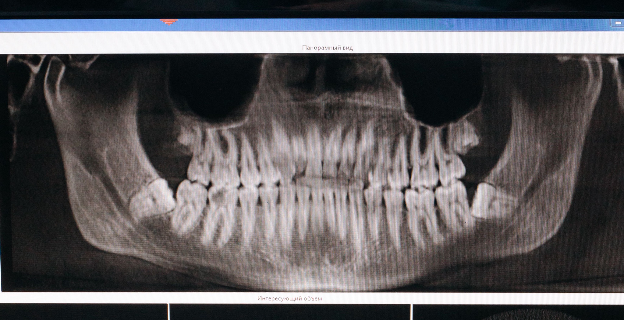
We integrated a regulator-cleared caries and bone-loss detection engine into the imaging viewer the group already used, so every bitewing and panoramic radiograph now renders with colour-coded overlays. Around it we built the working routine: a review queue where a clinician signs off every case, chairside views for talking patients through findings, and a group dashboard covering all 40 offices. Early-stage caries were flagged on 12% more bitewings than the pre-rollout audit baseline, second opinions now come back in under 4 hours, and treatment acceptance for recommended restorative work rose from 46% to 58%.
The Problem
Inconsistent radiograph diagnostics
The group had grown to 40 offices across the US Southeast, and radiograph reading practice had grown with it: office by office, clinician by clinician. Audit work showed early-stage caries and bone loss being flagged at very different rates from one office to the next. The image coming off the sensor was the same kind of evidence everywhere; what happened to it afterwards was not.
When a clinician wanted another set of eyes on a radiograph, the route was email. Images were attached, forwarded and queried in inbox threads, and answers took days to arrive. By the time a second opinion landed, the patient had often already left with a provisional plan, and the treatment conversation had to be reopened from scratch.
There was also no standard workflow for reviewing radiographs before treatment planning. Each office decided for itself when images were reviewed, by whom and against what threshold, which left group leadership unable to compare offices meaningfully or to say with confidence that a patient would hear the same story in any of them.
Inconsistent flagging
Early-stage caries and bone loss were flagged at very different rates between offices, so the same radiograph could lead to different treatment conversations depending on location.
Second opinions by email
Cross-office consults ran through inbox threads, with images attached and forwarded by hand, and answers routinely took days rather than hours to come back.
No shared workflow
Every office had its own habits for when radiographs were reviewed, by whom and against what threshold, with nothing standardised at group level.
No group visibility
Leadership had no view connecting radiograph review to treatment decisions across offices, so variability between clinicians stayed invisible until an audit surfaced it.
What it was costing them
Diagnostic variability carried a quiet price. Early-stage findings missed in one office became larger treatments later, patients received different recommendations for the same picture depending on where they sat, and days-long second opinions stalled treatment planning. Meanwhile clinicians who wanted to compare notes had no shared reference point, and the group could not demonstrate a consistent standard of radiograph review to anyone who asked.
The Solution
AI detection in every viewer
We started inside the viewer the clinicians already used rather than adding another screen. A regulator-cleared caries and bone-loss detection engine was integrated into the group's existing imaging software, rendering colour-coded overlays on every bitewing and panoramic radiograph as it is opened. Nothing about capturing images changed; what changed is what every clinician sees when they look at one.
Around the engine we built the rollout workflow that makes the output usable. A review queue holds every AI-annotated case until a clinician signs it off, so the software proposes and the human decides. Chairside presentation views let clinicians walk patients through the overlays during the visit, turning the radiograph into something a patient can actually see and discuss.
Finally we gave the group a single view of itself: a dashboard tracking detection and treatment-acceptance rates across all 40 offices. Onboarding ran office by office against the same playbook, and the standardised review workflow was in place everywhere within 5 months, so leadership could watch consistency improve rather than take it on trust.
Key decisions
Integrate, do not replace
The detection engine renders inside the imaging viewer clinicians already use, so adoption required no new application, no separate login and no change to image capture.
Mandatory clinician sign-off
Every AI-annotated radiograph waits in a review queue until a clinician signs it off, keeping the treatment decision with the human and the software in a supporting role.
Overlays built for chairside
Colour-coded overlays double as a patient communication tool: chairside presentation views let clinicians show patients what was found rather than describe it from memory.
Measure the rollout
A group-level dashboard tracks detection and treatment-acceptance rates across all 40 offices, so consistency became something leadership could observe instead of assume.
One playbook per office
Onboarding followed the same standardised workflow in every office, which is how the whole group moved onto the new routine within 5 months.
Measurable Impact
What changed after launch
The numbers moved in the direction the group had been chasing. Early-stage caries were flagged on 12% more bitewing radiographs than the pre-rollout audit baseline, and second opinions that once crawled through email now come back through the shared review queue in under 4 hours. All 40 offices were onboarded to the standardised review workflow within 5 months.
The chairside overlays changed the treatment conversation as much as the reading routine. With patients able to see the findings on their own radiographs, treatment acceptance for recommended restorative work rose from 46% to 58%. And because every office now follows the same review workflow, the group can finally talk about one consistent standard of radiograph review rather than 40 local versions of one.
Diagnostic consistency
Flagging rates varied widely from office to office
Early-stage caries flagged on 12% more bitewings than baseline
Second opinions
Email threads with answers taking days
Shared review queue turnaround under 4 hours
Review workflow
No standard process before treatment planning
One standardised workflow across all 40 offices
Treatment acceptance
46% acceptance for recommended restorative work
58% acceptance with chairside overlay conversations
Headline results
Early-stage caries flagged on 12% more bitewing radiographs versus the pre-rollout audit baseline
All 40 offices onboarded to a standardised radiograph review workflow within 5 months
Cross-office second-opinion turnaround cut from days to under 4 hours via the shared review queue
Treatment acceptance for recommended restorative work rose from 46% to 58% with chairside overlays
Tech & Tools Used
What powered the build
Every tool below earned its place in this engagement. Here is the part each one played.

Python (FastAPI)
Serves the integration layer between the imaging viewer and the detection engine, handling analysis requests and returning overlay data to the review workflow.

PyTorch
Runs the detection models inside the regulator-cleared engine, producing the caries and bone-loss findings that the colour-coded overlays render for clinicians.

ONNX Runtime
Executes the exported detection models in production, keeping inference fast and consistent so overlays are ready by the time a radiograph opens in the viewer.
Orthanc (DICOM)
Acts as the DICOM layer that receives bitewing and panoramic radiographs from office imaging systems and hands them to the analysis pipeline.

PostgreSQL
Stores findings, sign-off records and review-queue state, giving every AI-annotated case a durable audit trail from detection through to clinician decision.

Next.js
Powers the review queue, chairside presentation views and the group dashboard, all delivered in the browser so offices needed no new software installs.

Redis
Queues analysis jobs and caches worklist state so the review queue stays responsive during the morning rush of newly captured radiographs.

Docker
Packages the detection service and its dependencies into identical containers, so every environment runs the same stack from pilot through full rollout.
AWS S3 + ECS
Object storage holds the radiograph archive and overlay artefacts while the container service runs the detection stack, scaling with imaging volume across the group.
Ready to Build your Dental Healthcare Business with Image Segmentation & Analysis
Ask Byte
Ask Byte
Typically replies instantly
just Now
Hi! I'm OrganByte's assistant. How can I help you today?
AI-generated content may be incorrect